Sunday 30 March 2014

Laser Doppler Flowmetry


Laser Doppler Flowmetry 


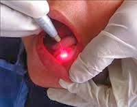
This is a non invasive, objective, painless, semi-quantitative method, which is more reliable in measuring the blood flow to the pulp. As it doesn’t cause any noxious stimuli, apprehensive or distressed, patients accept it more readily than the current methods to assess the pulp vitality. Laser light is transmitted to the pulp by means of a fiber optic probe. Laser Doppler flowmetry uses Helium Neon (HeNe) and Gallium Aluminum ( Ga AlAs) as semiconductor diode lasers at a power of 1 to 2 mW. The wave length of the HeNe laser is 632.8nm and that of the semiconductor diode laser is 780 to 820nm


The scattered light from the moving red blood cells in the circulation will be frequency-shifted, while those from the static tissues remain unshifted. The reflected light composed of Doppler shifted and unshifted light is returned by the afferent fibers and a signal is produced. This technique can be successfully employed for estimating the vitality of the pulp in both adults and children. The tooth to be checked should be isolated. The closer the probe is positioned to the gingival margins, the higher the signal output because of the greater volume of the pulp tissue .At the same time, the potential gingival contamination is also higher. The ideal position to place the probe is 2 to 3 mm from the gingival margin


Different ranges of band width can be set to filter the reflected signal, with a wider frequency being more sensitive to the moving red blood cells with a wider range of velocity .Theoretically, a wider bandwidth such as 15kHz is preferred, but in case of pulp vitality testing, a much narrower 3 kHz bandwidth may be ideal The end of the LDF which contacts the tooth contains both sending and receiving optic fibers, with one of the configuration being one source and two detectors in a triangular arrangement at the probe end Calibration of the probes is important to ensure accurate readings .


The larger the optical fiber separation distance on the probe, the higher the signal output as a larger surface area is covered, and also there is potentially a higher chance of blood flow signal contamination of the non pulp sources. To date, the 0.5mm or 0.25mm separation distances seem to be preferred in experiments. Due to the pulsatile nature of the blood flow, many studies.... have observed that the LDF recordings in the teeth with an intact pulp blood flow have rhythmic fluctuations or oscillations. In teeth without pulp blood flow, however, usually only irregular fluctuations can be observed in contrast to the concurrent ECG readings.


Tuesday 25 March 2014

ROLE OF LASERS IN ENDODONTIC ROOT CANAL THERAPY !


Decontaminating a root canal consists of killing bacteria, vaporizing dead and diseased tissue, and cleansing the canal of the residue. 


Nearly 20 years ago, lasers were proposed as a decontamination aid. As a bactericidal agent, the near infrared wavelengths generate fatal structural modifications in bacteria cells. The primary damage takes place in the cell wall, causing an alteration of the osmotic gradient, leading to swelling and cellular death. The highest bactericidal effect is on E. coli (Gram-negative), and E. faecalis (Gram-positive). In addition to killing bacteria, the near infrared wavelengths can ablate the root canal biofilm, which comprises the primary bacterial substrate. Biofilm constituents include the smear layer, debris, residual pulp and collagen fibers.


The near infrared wavelengths are well suited for decontamination in that they balance absorption and penetration with respect to sensitive tissues. On one hand, hard dentinal tissues do not readily absorb the near infrared wavelengths; diode irradiation therefore has no ablative effect on dentinal surfaces. The risk of damaging root canal tissues is greatly diminished. The trade off with low absorption is increased penetration.

 In light of being absorbed, diode irradiation is able penetrate the dentinal walls at subablative levels. Light at the 810nm wavelength can penetrate up to 750µ; the 1064nm wavelength can penetrate up to 1mm. Penetration promotes decontamination on deeper dentine layers. Medium infrared lasers can also be used in root canal decontamination. There are advantages and disadvantages.*

Diode laser energy is able to activate chemical irrigants to increase their chelating capacity. This results in more complete removal of the smear layer. The most common irrigants are sodium hypoclorate (NaOCl) and calcium hydroxide (Ca(OH2)), the latter mixed with propylene glycol and camphorated paramonochlorophenol. For example, Benedicenti et al. (2007) show the efficiency of diode laser decontamination in combination with 17% EDTA, 10% citric acid and 5.25% sodium hypochlorite. Chelating helps laser light to penetrate into the dentinal walls promote decontamination (ibid). Laser-activated irrigation has been shown to be statistically more effective in removing debris and the smear layer in root canals compared with traditional techniques and ultrasound.


Diode laser root canal decontamination is usually done with the smallest available optical fiber diameter: 200µm. The endo fiber fits well in most prepared root canals; it is flexible enough to penetrate all but the most severe canal curvatures. The 200µm fiber usually allows insertion to within 1mm of the canal apex. As the fiber emits from its distal end, the target tissue lies directly in front of the tip. A fiber-delivered diode beam diverges 15-22° upon exiting the tip. The emission heats an area slightly larger than 200µm, but the beam does not fully irradiate all canal walls. In decontamination therapy it is therefore recommended to bring the tip into the apical area, begin emission, and then withdraw the tip with a circular motion. With this simple method, canal walls can be effectively irradiated.


Tuesday 18 March 2014

5 LIFE CHANGING REASONS TO CONSIDER LASER HAIR REMOVAL !!!

Our relationship with hair is complicated. It's one of the few things on our bodies we can easily segment into two categories: wanted and not-wanted. How many other things on your body do you continually have to get rid of? There aren't many.Laser Hair Removal is life changing.

           Here are five of the top ways laser hair removal will change your life. 


                                        Get Smoother Skin


Laser hair removal removes hair at the root. The result is there is no more stubble, just amazingly smooth skin.         


                                          Eliminate Ingrown Hairs        


In-grown hairs are the worst! They're painful, they irritate the skin, and in worst case scenarios they can actually require surgery to remove. Eliminate the problem and take away the worry with laser hair removal. Because the laser removes hair at the root, there's no chance of ever seeing an ingrown hair. It's all gain, no pain.


                                          Gain Self-Confidence


As we get older, we all get hair in unwanted places.. With laser hair removal, you can remove unwanted hair permanently. You'll wake up looking clean, smooth, and beautiful. You'll never worry about those problem areas again!


                                          Save Your Time


Let's get real for a minute. Have you ever had more than one person needing to take a shower in the morning around the same time? Enough so that you routinely run out of hot water? Especially if you have kids, it happens all the time. If you've been there, you know that rushing through your morning shave is a recipe for cuts, missed spots, and the last person showering still ends up with cold water!
Eliminate all of that pain and hassle while leaving enough hot water for the last morning shower with laser hair removal.


That 5-10 minutes you save the 15-20 times you shave each month really ads up. Over 30 years, you can easily save more than 1200 hours. That's 50 DAYS of non-stop continuous shaving. Compare that with laser hair removal treatments, which total just a handful of hours and that unwanted hair is gone permanently.


Friday 14 February 2014

7 Ways Lasers Can Lead to More Patients


1  Less Referrals to Specialists – Soft-tissue lasers not only give practitioners the ability to precisely complete routine procedures performed in general practice, but also many procedures they previously would have referred to specialists.They are particularly useful for gingivectomies to facilitate caries removal and enhance a smile’s esthetics in a minimally invasive way.
3  Easier Periodontal Treatment – Lasers can be used for full periodontal therapy, including removing bacteria in patients with periodontal disease. According to Dr. Sam Low, a leading expert on technology for treating periodontal disease, “In addition to the long-standing benefits of diode lasers destroying bacteria in the periodontal pocket, there are exciting new dual wavelength protocols that are showing unprecedented tissue response and results for the patient. It is an exciting time for lasers in dentistry and the treatment of periodontal disease.”
4  Quick and Easy Cavity Treatment … that even younger patients won’t mind - One of the most promising aspects of laser dentistry is the opportunity to do more with less anes- thetic, less damage and less pain.
5 Better Impressions – When used to create a trough around prepared teeth, lasers can improve the quality of daily impression taking, resulting in impressions that are cleaner and more accurate.
6 Whitening and Cosmetic Dentistry Opportunities – Dentists can use a laser to improve the look of their patients’ teeth by removing part of the tissue to make the teeth match in size. PLUS, studies have shown that Biolase’s EPIC™ can perform a full- mouth, in-office whitening procedure with an average of 4-6 shades in only 20 minutes of treatment time. With LaserGel20 whitening gel kits available in five-packs, it’s a reliable, affordable treatment that can generate a high return on your investment.


7 Generate Buzz Around Your Practice –  With the persistent presence of social media in your patients’ lives, lasers like the WaterLase, which offer a transformational experience for your patients, can generate viral marketing about your practice and the type of dentistry you bring to your community.


Friday 7 February 2014

Occupational hazards to dental staff !!!

Dental professionals are predisposed to a number of occupational hazards. These include exposure to infections (including Human Immunodeficiency Virus and viral hepatitis); percutaneous exposure incidents, dental materials, radiation, and noise; musculoskeletal disorders; psychological problems and dermatitis; respiratory disorders; and eye insults.

Basically, for any infection control strategies, dentists should be aware of individual protective measures and appropriate sterilization or other high-level disinfection utilities.
Dentistry is considered by the practitioners and most of the public as being extremely hazardous. The hazards include the following:

Infectious Hazards: Needles and other sharp objects spatter, and aerosols can transmit viral infections such as life-threatening infections such as acquired immunodeficiency syndrome and hepatitis B. Bacterial infections also play an important role. The leading causes that should be concerned are syphilis and tuberculosis. 

Psychological Hazards: Stress is the leading psychological condition that occurs in the dental profession. Many studies implicate that dentists perceive their profession as more stressful than other jobs. Negative picture induced by the media of dentistry as a profession filled with dangers may be the leading causes.


Allergic Reactions: Gloves containing latex are the main causes of the allergic skin irritation, but dental materials, detergents, lubricating oils, solvents, and X-ray processing chemicals could lead to an allergic skin reaction. 


Physical Hazards: These include musculoskeletal complications which have direct relation to dentistry procedure, like postural situations that may increase the risk of twisting and contorting the body, varicose, etc.  


Mercury Health Hazard: It has been proved that high mercury vapor high dose exposure can lead to biological and neurological insults. Sealed amalgam capsules use with lower mercury level, water irrigation and high suction, good ventilation and proper collection, and discarding of amalgam have substantially diminished the mercury dangers. 


Ionizing Radiation: Taking X-ray machines in the dental office predispose dentists to suffer from ionizing radiation.


Non-Ionizing Radiation: This has recently become a concern since the use of composites and other resins, next to the use of lasers in dentistry procedures, which has added another potential hazard to eye and other tissues that may be directly exposed. 


Anesthetic Gases in the Dental Office: Using nitrous oxide gas regularly over an extended period of time may contain hazard.


Concerning prevention, the global literature focuses strictly on control of infections and appropriate management of potentially infected materials, owing to the high profile of dentistry regarding infection transmission. Barrier utilities such as gloves, masks, protective eye wear, high power suction, and good ventilation reduce aerosols and vapor hazards. Hypoallergenic non-latex gloves can decline latex allergy. Lead aprons, periodic maintenance of the X-ray machine and radiation level sensors deal with radiation dangers.

So, it is important for dentists to remain constantly up-to-date about measures on how to deal with newer strategies and dental materials, and implicates the need for special medical care for this professional group.


Friday 31 January 2014

WHY ARE WISDOM TEETH/3RD MOLARS OFTEN EXTRACTED BY DENTISTS

Wisdom Tooth or the 3rd Molar is the most commonly Extracted tooth due to pathological reasons by Dentists. Wisdom Teeth or 3rd molar teeth are being considered as vestigial parts of the human body as with evolution over decades it is seen that in some people 3rd molars are completely absent not even tooth buds are seen, and in others the jaw size decreasing constantly is the main reason for the lack of space for the 3rd Molars to erupt into Occlusion and Leading to Impaction. 


There are various steps in Local Examination to diagnose Impaction and the type of Surgery required to remove it. For example Wharf’s Assessment of Impacted 3rd Molar. 

WHY A DENTIST PREFERS REMOVAL OF 3RD MOLAR & ASKS THE PATIENT TO GET IT EXTRACTED.

REASONS FOR 3RD MOLAR EXTRACTION:

Most Commonly Impacted Tooth:

We all know that 3rd molars are the most commonly impacted teeth in the oral cavity after them comes the canines, but because of impaction the other teeth in the oral cavity are affected mostly 2nd Molar which is adjacent and due to the position of the 3rd molar it is most commonly affected. If there is Mesial Impaction of 3rd Molar there is a trench formed between the crowns of 3rd & 2nd molar which will lead to food accumulation and if there is horizontal impaction the crown of 3rd molar will impinge on the root of 2nd molar leading to resorption of the root. Hence for this main reason 3rd molar is the most extracted tooth in the Oral Cavity.

Lack of Space in the Dental Arch:
Due to constant evolutionary changes to living being the change in living conditions and the lack of requirement of that extra tooth to chew and to get good appearance the Jaw Size of Humans is decreasing and this is leading to loss of Space in jaw for the 3rd molar to erupt which takes place after the growth stage is finished in Boys/Girls.


Lack of space due to decrease in Jaw Size is the major reason for impaction of 3rd molar which in turn leads  to Extraction of the impacted 3rd Molar.

Unwanted Location of the tooth:
Due to lack of space and the tooth having no place to go the tooth is seen to get stuck in various unwanted locations which are classified as Mesial, Distal, Horizontal, vertical Impaction and this is the cause of various conditions like caries, cysts, Pericoronities etc and the only solution is removal of this harmful tooth. Sometimes the root of Impacted 3rd molar most commonly impinges on the inferior or Dental Alveolar Nerve Canal, so Relation of 3rd molar to Dental/Inf alveolar nerve canal is important in diagnosing & deciding the treatment plan. 


Difficult to Clean & Prevent caries:
Due to the location of 3rd Molar in the far end of the oral cavity & at many times because of its odd locations makes it very difficult to clean and remove Food & Debris from accumulating which will lead to caries to the Impacted tooth & along with it the adjacent 2nd Molar Caries is seen in both 3rd Molar & the distal aspect of 2nd Molar, so to protect the2nd molar removal of 3rd Molar is compulsory.

Common reason for Pericoronitis:
Pericoronitis is the most common complain in 3rd molar impactions and the most common reason why people come to know that they have impacted tooth and visit the dentist. Pericoronitis is nothing but inflammation of the soft tissue covering the crown of the Impacted 3rd molar because it constantly gets impinged between the 3rd & 2nd molar crowns. 
  

Friday 24 January 2014

What is Root Canal Treatment and Why Would You Want It?

Why Teeth Need Root Canals


Overall if we take the cases, approx. 85%-90% of the time a patient needs a root canal is due to dental decay (a.k.a. cavities).  In these cases, the bacteria from the decay enter the nerve of the tooth, resulting in pain for the patient.  This should be very intuitive: you have live nerve tissue that is normally sequestered from the outside environment which is now suddenly exposed to nasty bacteria.  Of course it is going to hurt! The other teeth needing root canals are primarily due to other factors such as trauma, cracking, and resorption.
                                                                                                                                                                                                                                            Root Canal Picture

Why Patients Associate Pain with Root Canals

A woman in pain needing a root canal. She will associate this pain with the actual root canal procedure.


This scenario plays out fairly frequently.  A patient with extreme tooth pain.  The pain is often described as “unbearable” or “the worst pain I've ever had” or sometimes even “worse than childbirth.”  An x-ray is taken and we perform a limited exam. We observe dental decay into the nerve of their tooth.  We discuss the findings with the patient and recommend a root canal. The patient consents.


                                                                                                                        Woman with a toothache
                                                                                                                         needs a root canal


We place topical anesthetic over the injection site and assure the patient that the pain will soon be gone. We then administer the local anesthetic with the patient feeling little to no sensation of the needle.  Within just a few minutes, they are profoundly numb, and their pain is gone!  We then perform the root canal procedure, removing the bacteria in the tooth, and then filling the roots of the tooth.  The procedure now done, the patient goes home, the numbing wears off, and the pain is still gone.  
And it is gone for good and they still have their tooth!!!!


Years later, the patient will then recount his/her experience, and say:

Yeah, I had a root canal at the dentist. It was the worst pain I have ever experienced!

This happens all too often. The patient confuses the pain that caused him to need the root canal with the pain of the procedure.  And every person who listens to this story then thinks that root canals hurt.  And thus, the myth lives on!

So remember:
  • Root canals do not cause pain, they relieve it.
  • Root canals allow you to keep a much compromised tooth.
  • There are no substitutes for your own natural teeth.


Thursday 16 January 2014

AC Vs. DC X-Ray Generators....


With the rise of digital radiography, equipment manufacturers have been releasing DC x-ray machines and touting them as the AC best x-ray AC machine for digital radiography.

AC Best AC is of course, often a sales strategy. So what is a DC x-ray, and is it really better?


AC x-ray machines use an alternating current to drive the tube head to produce x-rays. This is usually the same 120v AC supply that powers our home; the voltage alternates from +60 to -60 volts, giving 120v of potential across two wires. The tube head produces x-rays for only about 1/2 of the total exposure time, as radiation is produced only during the positive alternation of the AC waveform. This method has been used to produce dental x-rays since their inception. It is a relatively simple technology and works very well.

DC x-rays are actually high frequency AC machines – rather than a 60 Hz waveform like an AC x-ray, they produce a very high frequency (often around 70 kHz) waveform, and only produce positive pulses. The end result is a much more constant output of radiation from the tube head.


What this means to the user is that during very short exposure times, the DC x-ray will produce more consistent radiation than the AC unit, by as much as 1/3 on a .04 sec exposure. As a result, images taken with a DC x-ray tend to be more consistent from image to image. However, this advantage disappears rapidly as the exposure time increases. Someone taking standard film-based bite wings at .25 seconds would not see the difference.

DC x-ray machines can provide more consistent images from low exposure time settings, but there is no need to replace x-ray machines just to go digital. The AC x-ray generator that currently hangs on your wall is more than capable of producing high quality, diagnostic images, even with digital radiography. However, if you are building a new practice or looking to replace an x-ray machine in the near future, it is worth taking a closer look at a DC unit.


If you are looking dental x ray machines check the listings of new dental x ray machines on our site.

Friday 10 January 2014

Top 10 Dental Product Categories of 2013 !!!

We begin our year in review with a high level view at the most popular dental product categories of 2013.

The categories on this list reflect a mix of the most exciting technologies on their way to changing the industry and the backbone technologies already powering dental practices. While cutting edge developments and new breakthroughs might get people excited, the top categories are a bit more grounded, showing the types of dental products and technologies that every practice needs, or is at least considering.

Now here are the Top 10 Dental Product Categories of 2013

10. Dental Loupes – Magnification is important, and more and more clinicians are realizing that when they can see with more detail, they can provide better care. If you and your team don’t use loupes when you practice, now is a great time to consider them. 


9. Core Build UpMaterials – There’s always an outlier on these lists, and this year it’s core build up materials. While not the most exciting, even in the rapidly developing world of dental materials, it seems 2013 was a year you and your colleagues were interested in your options for completing endodontic cases.


8. Practice Management Software – This is a product that is truly the heart of a dental practice. The software available to manage your patient data continues to grow and evolve with new integrated marketing features as well as the exciting possibilities of cloud software replacing the current server-based software model.

7. Dental Autoclaves– With some high profile breaches of protocol and the resulting serious consequences making the news this year, it’s not a surprise to see dental autoclaves on this list. This is a product every practice must have, and finding a sterilizer that fits your practice’s needs is the first step in setting up a solid infection control system.


6. Dental LED CuringLights – Dental materials continue to improve and the technology required to properly cure everything from adhesives to composites is changing just as quickly. 2013 saw the launch of some notable new curing lights, and that likely helped keep interest in these products high. 


5. Cone Beam 3DImaging – Imaging is critical to modern dentistry and many of the latest imaging technologies made this list. Cone beam CT is growing in prominence as 3D patient information makes diagnosis and treatment planning easier. While the cost of these systems is high, many dentists are discovering they can realize a return on the investment if they have a solid plan to use the new imaging technology.


4. Digital PanoramicX-Ray – Another imaging technology ready to help clinicians improve their diagnosis and treatment planning, digital panoramic systems remain a solid part of the digital imaging landscape. For many practices digital panoramic systems represent a more cost effective way to improve the imaging protocol.


3. Dental Diode Lasers – Used for a range of soft tissue and cosmetic dental applications, the diode laser continues to gain traction in operatories around the world. These lasers make it easier to provide minimally invasive care in a number of situations. If you’re not using one now, it might be time to give them a look. The technology keeps improving and diode lasers are easier to use than ever. 


2. Intraoral Cameras – Another imaging technology that is a popular addition to numerous dental practices, the intraoral camera is the best way to show patients what you see. When it comes to case acceptance, the intraoral camera might be the most powerful technology out there as your patients are likely to understand a picture taken from inside their mouth more readily than your description of what’s happening in there.

1. Digital Dental X-Ray Sensors – The most popular product category of 2013 is the intraoral x-ray sensor, and this should be no surprise. Digital radiography is far past the tipping point and on its way to not just being the standard of care, but the norm in dental practices everywhere. Patients are aware that digital x-rays mean less radiation exposure, and clinicians know the digital radiographs are faster to take, easier to store and better for diagnostics.

Friday 3 January 2014

Chromo therapy Light Can Benefit Our Well-Being


What is Chromo therapy?
Chromo therapy or Color Therapy healing is the science that uses the 7 colors of the spectrum to improve our mental well-being and the health of our bodies. The 7 colors have the ability to affect the vibrations of the body in the frequencies that affect health, welfare and harmony. Color therapy has been used in Asia for a thousand years.

Chromo therapy is used for a variety of conditions including depression, anxiety, stress, fatigue. pain. cramps, headache,migraine headache, diabetes, high blood pressure, asthma, cough, and many other conditions.

Benefits of Chromo therapy Colors...
In addition to the 7 main colors of the spectrum, there are 5 additional colors that are beneficial to our bodies:


How does it work?
Chromo therapy practitioners believe that color can be used to correct energy imbalances which are the cause of disease. A chromo therapist applies specific colors or lights to specific points on the body called “chakras.” Different colors have different effects. For example, red is thought to increase pulse rate, blood pressure, and breathing rate. Therefore, it is often used for circulatory conditions. Blue is believed to cause relaxation and calm. Therefore, blue is used for headaches, pain, cramping, stress, and other conditions.


What are the Chakras that are used in Chromo therapy?
Chakras are the energy points that are located in the aura, the field of energetic vibrations that exists in every human body. Chakras have the shape of a wheel or vortex. In the human body they have an average diameter of 5 to 6 centimeters (2").


Connection of Chakra Colors to Illness...

The 7 main chakra colors are commonly defined as...